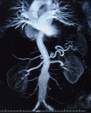
Management of trauma demands experienced and multidisciplinary team work in which radiologist should be involved in both diagnosis and treatment. On one hand scanning the entire body is key in identifying lesions and on the other hand endovascular treatment (stents and embolization), performed by interventional radiologists.

At the scene of the accident rescue teams identify and correct deficiencies and achieve the vital balance. Considerable progress has been made in the medicalization of rescue and transport of the wounded, reducing morbidity and mortality. Equally decisive progress has been made in the emergency department, benefiting from a coordinated multidisciplinary care generally guided by an anesthesiologist.
Everything must be perfectly organized because every minute counts. The highway trauma victims are often young subjects who were previously healthy. Everything must be done to save the patients in such a manner if possible to leave the minimum of scars so that may resume a normal social and professional life.
Once the vital functions are stabilized, a CT scan of the whole body is the ideal investigation method. In 3-4 minutes brain and abdomino-pelvic lesions can be reviled and thus prioritizing therapeutic strategies. The multidisciplinary team including radiologists, interventional radiologists, anesthesiologists, vascular surgeons, orthopedists, neurosurgeons, decide the therapeutic approach. This involves having a suitable technical platform and all of these specialists available round the clock.
The role of the CT and angiography in emergencies:
Traffic accidents may be responsible for damage of large arteries located in the chest or abdomen, including the aorta, iliac or sub-clavia arteries, causing massive bleeding sometimes. These lesions usually occur in the context of poly-trauma and are immediately lethal to 80% of patients.
Interventional radiologists can offer these patients a chance to fight! By being able to see the bleeding lesions, they can treat all arterial territories. To stop the bleeding radiologists use different techinques for hemostasis. Embolization can occlude the artery, either permanently or temporarily. The other procedure is to establish a covered stent to ensure the restoration of the continuity of the injured vessel, as it’s done in the context of cerebral aneurisms.

For the full trauma of abdominal organs (kidney, spleen, liver), the classification of the American Association for Surgery of Trauma refers to define treatment options. It distributes the visceral abdominal trauma in 5 stages of increasing severity. Embolization is indicated in patients with hemo-dynamically stable or controlled hemorrhagic shock in the presence of the active scanner leakage of contrast, false aneurysms, arteriovenous fistulas and depending on the severity.
Advances in materials
Regarding the renal trauma, depending on the type of injury, several interventional radiology techniques can be proposed: stents for truncal arterial rupture, embolization of acute hemostasis, secondary treatment of arterovenous fistula tracts.
For the spleen, ablation remains the rule for all patients in uncontrolled hemorrhagic shock. In other cases, embolization is indicated because it can retain a portion of parenchyma and therefore the function of the organ.
For the liver arterial embolization.
Pelvic fractures, they are markers of high velocity trauma. These may be accompanied by bleeding complications due to arterial bleeding, venous bleeding ore bone. Endovascular embolization should be preferred instead of the surgical approach.
All these procedures have benefited from advances in materials and devices. Depending on the type and location of lesions, the interventional radiologist chooses absorbable product that provides temporary occlusion and allows, after 3 to 4 weeks, the artery become permeable, or a non-resorbable. The range has also expanded stents and radiologists have devices of any diameter and any size.
Source of the article hereÂ


